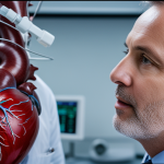
Immediate strategies to minimise radiation exposure in UK medical imaging
UK radiographers focus heavily on radiation reduction techniques to enhance patient safety. One primary approach is adopting internationally recognised and UK-specific protocols designed for dose reduction. These protocols incorporate the principles of justification and optimisation, ensuring every imaging procedure is necessary and performed with the lowest possible radiation dose.
In practice, justification means each scan is carefully evaluated against potential benefits and harms. Optimisation involves tailoring equipment settings and scanning parameters, balancing image quality with minimal radiation. Integrating these principles into the daily workflow helps radiographers consistently reduce unnecessary exposure.
Additional reading : Refurbished refraction units for optometry: quality and affordability
Additionally, practical strategies include positioning patients correctly, employing shielding devices where applicable, and selecting the most appropriate imaging modality. UK radiographers continuously adjust techniques based on patient size, clinical indication, and previous imaging history. These combined efforts form a proactive framework that significantly diminishes radiation risks while maintaining diagnostic accuracy, reflecting a patient-centred commitment embedded in UK healthcare standards.
Innovative imaging technologies advancing dose reduction
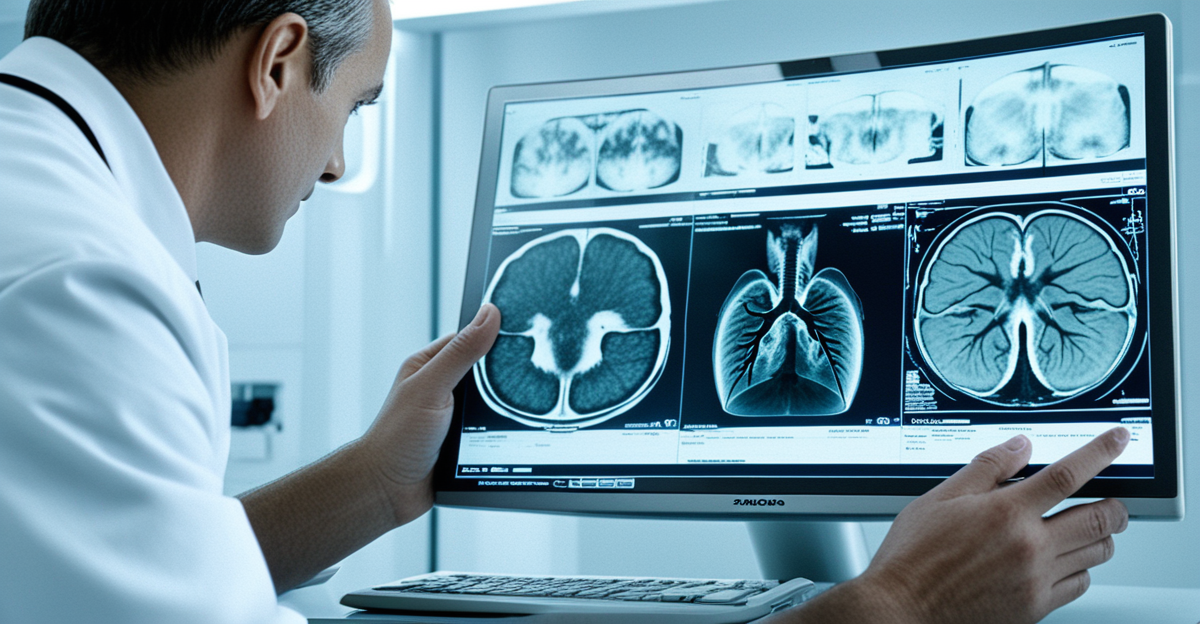
Advancements in medical imaging technology have significantly empowered UK radiographers to enhance radiation reduction techniques while maintaining diagnostic quality. The adoption of low-dose equipment, such as digital radiography systems, allows for detailed images at much lower radiation levels compared to traditional film methods. This shift is critical for improving patient safety by limiting exposure without compromising results.
In the same genre : Expert techniques for uk podiatrists: mastering diabetic foot ulcer care
UK hospitals increasingly implement iterative reconstruction CT techniques. These algorithms reconstruct high-quality images from fewer X-ray projections, reducing the dose required per scan. Combined with dose-tracking software, these technologies provide real-time monitoring and feedback, enabling radiographers to adjust exposure on the spot.
Moreover, AI-powered exposure modulation tailors radiation doses to individual patients based on size, anatomy, and prior imaging, demonstrating a precision approach aligned with optimisation principles. These innovations exemplify how radiology advancements directly support UK radiographers in advancing radiation reduction techniques, contributing to safer, more effective imaging practices.
Education and ongoing training for UK radiographers in radiation safety
Continuous radiographer training is essential to uphold high standards in radiation reduction techniques and ensure patient safety. UK radiographers undergo mandatory professional development focused on the ALARA (As Low As Reasonably Achievable) principle. This training emphasises dose optimisation, reinforcing knowledge of how to minimise exposure without compromising image quality.
Practical workshops and simulation exercises play a crucial role, allowing radiographers to apply radiation safety concepts in realistic scenarios. These hands-on experiences improve skills in selecting the proper imaging parameters and in positioning patients optimally—both vital for reducing unnecessary radiation.
Peer-review practices contribute by facilitating feedback among UK radiographers, encouraging continuous learning and adherence to best practices. Through regular updates on emerging technologies and evolving protocols, radiographers remain informed about cutting-edge radiation reduction techniques.
In summary, structured education and ongoing training empower UK radiographers to integrate radiation safety deeply into clinical workflows, reinforcing their commitment to patient safety with every imaging procedure.
Real-world examples and case studies from UK radiography departments
UK radiography departments provide clear case studies demonstrating successful implementation of radiation reduction techniques that enhance patient safety. For instance, several NHS Trusts have integrated patient shielding protocols, such as lead aprons and thyroid collars, into routine practice. This straightforward approach significantly reduces scatter radiation and protects vulnerable tissues during diagnostic imaging.
Another example highlights the impact of targeted radiographer training programs. Following comprehensive staff education on dose optimisation and positioning, some departments reported measurable declines in radiation doses without compromising image quality. These best practices reflect a commitment to continual improvement and demonstrate how ongoing professional development translates into safer care.
Furthermore, multidisciplinary collaboration involving radiographers, medical physicists, and clinicians fosters a culture prioritising patient-centred dose reduction. This teamwork supports protocol refinement and audit, ensuring lessons from case studies shape wider clinical governance. By sharing outcome data and strategies, UK healthcare institutions can replicate successes and drive uniform standards in radiation safety.
In sum, these real-world examples confirm that well-implemented strategies and staff engagement combine effectively to reduce unnecessary radiation exposure across UK healthcare settings.
The role of guidelines and regulatory bodies in promoting safer imaging
UK radiographers rely heavily on robust imaging guidelines and regulation to ensure consistent application of radiation reduction techniques and promote patient safety. The Ionising Radiation (Medical Exposure) Regulations (IR(ME)R) provide a mandatory legal framework. IR(ME)R requires justification of every medical imaging exposure, ensuring scans are clinically warranted and optimised to minimise dose. This enforces accountability and standardisation in practice.
In addition, clinical governance structures embed these regulations into daily workflows. Through audit and quality assurance processes, adherence to safe imaging protocols is monitored and improved continually. Regulatory bodies collaborate closely with professional societies and Public Health England, updating guidelines based on emerging evidence and technology advancements.
These organisations also facilitate training resources supporting UK radiographers in maintaining compliance and using best practices. The integration of regulation into clinical governance ensures that radiation safety remains a priority across all UK healthcare settings. This comprehensive oversight supports radiographers in delivering safe, effective imaging while protecting patients from unnecessary radiation exposure.